
Heart Attacks Explained: Common and Less-Known Causes You Should Understand
Written by Dr Edward Leatham, Consultant Cardiologist: In this article, we shed light on various underlying factors that can lead to this cardiovascular emergency.
Providing independent clinical excellence since 2005
Posted on Saturday January 10, 2026 in VAT-TRAP

Tags: VAT, Metabolic Health, Visceral Fat, search website using Tags to find related stories.
If you truly want to understand what is happening inside your body — bone strength, skeletal muscle mass, and fat distribution — medical imaging is the only approach with sufficient accuracy to support best medical practice.
“Body composition” is not a cosmetic concept. It directly relates to outcomes that matter clinically and personally: hip fracture, vertebral compression and spinal collapse in later life, frailty, insulin resistance, cardiometabolic disease, and increasingly recognised risks such as dementia. Decisions based on imprecise measurements risk false reassurance, delayed intervention, or inappropriate therapy.
The challenge is that many commonly used metrics — weight, BMI, and consumer “smart” devices — create an illusion of precision. They provide numbers, but not necessarily truth. When it comes to guiding therapy, prevention, or long-term health planning, approximation is not enough.
In clinical medicine, meaningful body composition assessment rests on three pillars:
Each pillar predicts different outcomes. Bone density predicts fracture and loss of independence. Muscle mass predicts strength, recovery from illness, insulin sensitivity, and longevity. VAT predicts cardiometabolic risk far more powerfully than body weight alone.
Each pillar is best assessed using medical imaging.
Most home “body composition” scales use bioimpedance analysis. A small electrical current is passed through the body, and lean mass and fat mass are inferred using proprietary algorithms.
The limitations are substantial:
Even professional-grade systems do not carry sufficient accuracy to govern medical decision-making or therapy. At best, they provide directional trends.
Despite these limitations, body composition scales are a valuable part of our metabolic toolkit in patients with metabolically unhealthy visceral adipose tissue.
They are not used diagnostically. Instead, they are deployed as home-based tracking tools, providing frequent feedback between formal medical measurements.
When used consistently, they can:
Most importantly, they act as a powerful driver of behavioural change.
Sustained reduction in visceral adipose tissue requires behavioural change maintained over months. Behaviour change without feedback rarely succeeds.
This is why we combine home body composition scales with:
Together, these tools form a closed-loop system:
education → action → feedback → adjustment.
The aim is to gradually lower the defended set point for weight and VAT — the physiological goal that underpins durable metabolic improvement.
Dual-energy X-ray absorptiometry (DEXA) is the internationally recognised gold standard for assessing bone mineral density and diagnosing osteoporosis¹. Guidance from the International Society for Clinical Densitometry defines its clinical use and interpretation¹.
DEXA:
DEXA also provides robust estimates of lean mass, commonly used in sarcopenia assessment.
The radiation dose from DEXA is extremely low, typically well below that of a chest X-ray². This favourable safety profile allows direct access DEXA scanning in appropriate settings.
DEXA is excellent for bone and muscle assessment, but it is not the optimal tool for measuring visceral adipose tissue.
We offer direct access to DEXA, get in touch and one of our patient care coordinators will let you know pricing and availability.

Visceral adipose tissue is biologically active fat stored around abdominal organs. Unlike subcutaneous fat, VAT drives:
VAT is therefore one of the most important modifiable risk factors in modern medicine.
Importantly, VAT cannot be reliably inferred from weight, BMI, or appearance.
The most accurate methods for VAT measurement are MRI and CT.
MRI avoids ionising radiation but is slower and less accessible. CT is fast, widely available, and highly reproducible. Modern scanners allow substantial dose reduction compared with traditional abdominal CT³.
Large studies demonstrate that VAT measured from a single cross-sectional slice correlates strongly with total VAT volume⁴⁵.
Based on this evidence, at VCL/SCVC we use a very low-dose single-slice CT VAT scan.
Peer-reviewed radiology literature confirms that VAT can be accurately measured using a single CT slice, with an effective radiation dose of approximately 1 mSv — around 10 % of the dose of a standard abdominal CT⁷.
Using modern dose modulation, this provides:
This dose is approximately one quarter of that of a mammogram and far below historical abdominal CT exposures³,⁷.
GLP-1 receptor agonists have transformed cardiometabolic care. Their benefit extends beyond weight loss.
In the SELECT trial, semaglutide significantly reduced major adverse cardiovascular events in patients with established cardiovascular disease and overweight or obesity without diabetes⁶.
A highly plausible mechanism is preferential reduction in visceral adipose tissue, with downstream improvements in insulin resistance, lipid metabolism, and inflammation.
If cardiovascular risk reduction is the goal, VAT is the metric that matters.
Weight loss without muscle preservation increases frailty risk and undermines metabolic health.
Skeletal muscle underpins glucose disposal, metabolic flexibility, physical independence, and long-term weight maintenance. Our CT VAT report therefore also estimates skeletal muscle mass, allowing personalised protein targets:
Understandably, many patients are concerned when they hear the word CT scan. Much of that concern relates to experiences with full abdominal CT, which historically carried higher radiation doses.
The VAT scan we use is very different.
To put this into context:
This means the radiation exposure from a VAT scan is:
Crucially, the scan provides clinically meaningful information about visceral fat — one of the most important modifiable drivers of cardiometabolic risk — using a dose that is considered low and proportionate when appropriately justified.
We do not offer this scan indiscriminately. Every scan is:
In short, this is a targeted, low-dose investigation, designed to give high-value information while keeping radiation exposure as low as reasonably achievable.
All CT imaging must be clinically justified.
At SCVC and VCL, we operate a free pre-consultation pathway using a structured questionnaire. When criteria are met, scans can be arranged without formal clinic consultation.
If accuracy matters, medical imaging is essential.
Precision provides the anchor. Tracking provides the momentum. Together, they enable the sustained behavioural change required to lower the defended set point for weight and visceral adipose tissue.
If you are interested in finding out more, use our enquiry form and a patient care coordinator will get in touch.
Written by Dr Edward Leatham, Consultant Cardiologist: In this article, we shed light on various underlying factors that can lead to this cardiovascular emergency.
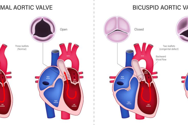
Aortic stenosis (AS) is a progressive and potentially life-threatening condition. It predominantly affects older adults and has significant implications for cardiovascular health, life expectancy, and quality of life. The incidence of AS is climbing because of increased life expectancy. Timely intervention, especially in symptomatic patients or those with progressing stenosis, is essential to optimise outcomes. This article explores the nuances of AS diagnosis, treatment decisions, and the importance of patient involvement in the care pathway.

LDL-C alone may miss important cardiovascular risk. This article explains why ApoB and small dense LDL often better reflect atherosclerotic burden, especially in metabolic disease and high visceral fat. It outlines practical lipid targets by risk category and shows how LDL-C:ApoB ratios can estimate dangerous particle patterns