
What techniques are out there to reduce glucose spikes?
This blog explores how individuals can conduct their own experiments to maintain a steady blood glucose and avoid potentially unhealthy glucose spikes above 7.8 mmol/L (140 mg/dL).
Providing independent clinical excellence since 2005
Posted on Wednesday September 17, 2025 in Naked Heart
An article written by Dr Edward Leatham, Consultant Cardiologist © 2025 E.Leatham
An AI audio construct is available as a podcast for this story below.
Tags: Mens Health, Visceral Fat, Coronary heart disease, search website using Tags to find related stories.
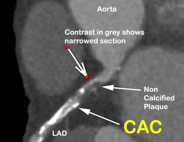
For over two decades, coronary artery calcium (CAC) scoring has been used as a quick, low-cost, low-radiation screening tool for identifying subclinical coronary artery disease. However, recent advances in cardiovascular imaging, growing understanding of plaque biology, and shifts in preventive cardiology have challenged its continued use in younger patients.
At our clinic, we no longer recommend CAC scoring as a screening test for men under 50 and women under 60. Here’s why.
CAC scoring detects calcified plaque, which represents only 10–20% of total coronary plaque volume in early and intermediate stages of disease¹. The remaining 80–90% of plaque is non-calcified, and this is particularly true in patients under 60. These early-stage plaques are vulnerable, inflammatory, and prone to rupture—even if they are not yet calcified.
We’ve seen too many patients in their 40s and 50s with significant non-calcified coronary plaque and a CAC score of 0. These individuals are often falsely reassured and do not get started on statins or lifestyle interventions when, in fact, they are at meaningful risk.
CAC does not measure stenosis or obstructive disease. It reflects the burden of calcification, not plaque volume, vulnerability, or flow-limiting disease. Numerous studies confirm that there is no direct correlation between CAC score and angiographic stenosis severity².
This limitation severely restricts the clinical utility of CAC in personalising risk or guiding therapy in younger adults.
Tracking CAC progression over time has little predictive value. An increase in CAC may reflect disease progression—or it may reflect healing, as statins and LDL lowering often promote plaque calcification as a stabilisation mechanism³. Repeating CAC scans does not provide actionable insight, nor does it alter clinical decisions in most cases⁴.
Because of this, serial CAC testing is not recommended by major guidelines⁵.
The effective radiation dose of a typical CAC scan is 0.8–1.5 mSv⁶—comparable to a mammogram, and lower than a standard CT angiogram. While this is low, any exposure to ionising radiation must be justified, especially in younger adults, where CAC is often absent and the scan is non-informative.
By contrast, a modern CT coronary angiogram (CTCA) can now be performed at 2.0–4.0 mSv with contemporary dose-reduction techniques⁷. For just a modest increase in radiation, CTCA provides vastly more information—identifying non-calcified plaque, assessing stenosis, and, increasingly, inflammation metrics like the **Fat Attenuation Index (FAI)**⁸.
One of the most exciting advances in cardiovascular imaging is the ability to detect coronary inflammation using the perivascular Fat Attenuation Index (FAI). Available as early as age 30, FAI provides predictive value beyond calcium or cholesterol, identifying at-risk individuals even in the absence of plaque⁹.
This imaging biomarker gives us a functional window into plaque activity, not just structure. When paired with plaque morphology and volume from CTCA, we can now offer personalised prevention strategies far earlier and more accurately than CAC alone ever could.
Ironically, one of the goals of lipid-lowering therapy is to stabilise plaque through calcification. Statins help convert soft, high-risk plaques into dense, calcified, low-risk structures¹⁰. As such, the presence of calcification may reflect prior healing—not current risk.
This further undermines the logic of relying on CAC in younger patients, whose plaques are often early-stage, lipid-rich, and uncalcified.
In 2025, with access to low-dose CT angiography, FAI analysis, and a deeper understanding of plaque biology, it is increasingly difficult to justify CAC scoring in younger individuals.
Given the modest but real radiation dose and the high false reassurance rate, continuing to offer CAC to younger patients may no longer be ethical, especially when more informative tools are available.
If the goal is early identification and prevention, CTCA with plaque and FAI analysis is the new standard—and CAC is best reserved for selected older patients where calcified burden more closely tracks with overall risk.
References
This blog explores how individuals can conduct their own experiments to maintain a steady blood glucose and avoid potentially unhealthy glucose spikes above 7.8 mmol/L (140 mg/dL).

Transoesophageal echocardiography before atrial fibrillation cardioversion prevents stroke by detecting hidden left atrial appendage clots. The greatest risk comes from atrial stunning after rhythm restoration, not existing thrombus. TOE enables safe early intervention.

Statins are a mind boggling biological 'ruse'. When this medication is taken orally, it causes liver cells to become deficient in essential cholesterol, so they respond by increasing the number of cell surface LDL receptors, thereby enhancing their capacity to extract LDL particles from the blood, significantly lowering circulating LDL levels. Statins remain the most effective and well-known way that doctors can lower LDL levels to prevent or treat coronary heart disease.