An article by Dr Edward Leatham, Consultant Cardiologist.
Coronary heart disease is a significant health concern that affects many individuals worldwide. It arises from the buildup of LDL (low-density lipoprotein) rich plaque within the walls of the coronary arteries. These arteries, crucial for supplying blood to the heart muscle, are relatively small, with diameters ranging from 2 to 4mm, and are located on the heart’s surface. Imaging these arteries in a beating heart presents unique challenges due to their movement and size.
Advancements in computed tomography (CT) technology have significantly improved our ability to visualize the coronary arteries. Noninvasive cardiac CT scans, enhanced with contrast agents injected into the arm’s vein, allow for detailed imaging and reconstruction of these arteries, facilitating the creation of an angiogram.
However, interpreting CT images can be complicated due to movement artifacts and the presence of calcific plaque, which can block x-rays and lead to overestimation of arterial blockage. This is particularly problematic because the decision to perform a coronary intervention, such as placing a stent, often hinges on whether a vessel is more than 60-65% blocked. Since atherosclerosis progresses slowly, determining the necessity of intervention for vessels with blockages in the 60-70% range can be challenging.
This uncertainty leads to a significant number of patients being referred for invasive angiography, a more sensitive test, which sometimes reveals no flow-limiting issues, rendering the invasive procedure unnecessary. Invasive angiography, requiring hospital admission, carries its risks, underscoring the need for more precise noninvasive diagnostic tools.
The development of a computational model by researchers at Stanford University marked a significant breakthrough in noninvasive coronary flow assessment. This model correlates with the catheterization laboratory’s gold-standard fractional flow reserve (FFR) test, providing a valuable tool for cardiologists.
Since 2016, cardiologists have been utilising the HeartFlow Analysis in conjunction with cardiac CT scans to enhance the accuracy and predictive value of noninvasive coronary imaging. This technology allows many patients to avoid unnecessary invasive angiography, reducing the associated risks and stress.
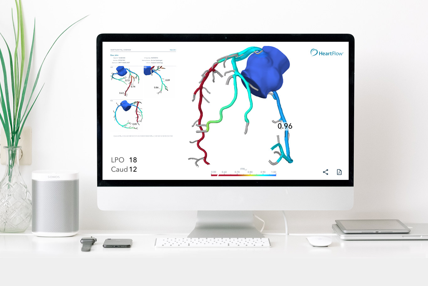
How Does CT FFR Work?
For the HeartFlow Analysis to be effective, high-quality CT images are necessary. These images are anonymized and uploaded to HeartFlow’s platform, where advanced algorithms, including deep learning, analyze the data to create a flow map. This map is color-coded and superimposed over a 3D model of the patient’s coronary arteries, providing a detailed visualization of blood flow and potential blockages.
The processed data is then returned to the reporting team and the referring cardiologist, who combine this information with their clinical expertise to make informed decisions regarding patient care.
This process is streamlined and enhances the diagnostic pathway for patients undergoing cardiac CT. By integrating HeartFlow Analysis, healthcare providers can assure patients of the reduced likelihood of unnecessary catheterization lab visits, focusing on interventions only when absolutely necessary.
In summary, CT FFR, particularly through the HeartFlow Analysis, represents a significant advance in the noninvasive diagnosis and management of coronary heart disease. It exemplifies the power of integrating cutting-edge technology with medical expertise to improve patient outcomes and reduce the need for invasive procedure.